baby chest x ray technique
Use gonadal shielding if possible. No evidence of pneumonia.

Radiologic Technique Charts Tech Nique Chart For Techniquetechnique Charts Radiologic Radiology Schools Radiology Tech X Ray
Immobilize infants arm above the head or use stockinette ace bandage tape or sandbags.

. Erect chest X-rays are taken at 180 cm. 901a a Use automatic exposure control 500 speed for chestabdomen else 400 speed at specified kVp when practical. Radiographs are also obtained to evaluate cause for dyspnea in the newborn.
This is partly due to modesty but also due to fear. Radiologists consider a chest X-ray to be of good quality when the trachea is centered and equidistant from the head of the clavicle on both sides the spine is visible as a transparent structure through the heart shadow and there is full inspiratory effort the right 6th rib is at the midpoint of the hemidiaphragm on that side. Full legfull spine imaging is performed at 180 cm using CR.
However all children are modest to some degree about having their genitals or backsides exposed after ages 4 to 5. AP and lateral chest x-ray demonstrates minimal peribronchial cuffing likely related to IV fluids. This approach allows you to assess the overall condition of the breast.
The anteroposterior AP diameter of the neonatal chest is almost as great as its transverse diameter giving the chest a cylindrical configuration. In Canada 33 hospitals were contacted initially as potential participants. Pediatric Chest Screen 70-80 DIGITAL OPTIMUM kVp Universal CR Technique Chart using a standard 21 LgM Part View kV mAs kV mAs kV mAs Abdomen AP Grid 85 10 -15 85 20 - 25 85 30 - 40 Ankle AP 70 18 70 2 70 25 Ankle Obl 70 16 70 18 70 22 Ankle Lat 70 15 70 16 70 2 Chest -Adult AP 400 - tt -72 85 2 - 25 85 32 - 4 90 5 - 64.
X-ray Imaging for Pediatrics. Patient Position Infant. The degree of rotation is best assessed by comparing the length of the anterior ribs visible on both sides.
Baby chest x ray technique. In most cases rib X-rays are performed in frontal and lateral projections. X-rays use a small amount of radiation about the same levels that occur naturally in the environment.
Lateral cervical spines are taken at 150 cm. The publication of this study and exposure chart could act. All distal extremity exposures are taken at 110115 cm SID.
Hospi-tals were excluded if they did not have an x-ray department a mobile x-ray machine or refused participation. As a benchmark for other medical imaging departments and to promote discussion on digital X. Chest AP erect in chair 180 cm.
Most neonatal chest X-rays are AP films unless the baby is made to lie prone Lucency of soft tissue shadow - darker the soft tissue more is the exposure Ease of visibility of retrocardiac vertebrae if the retrocardiac vertebrae are easily seen the film is over exposed Relative lucency of lung fields. The normal neonatal chest X-ray. In contrast most 12-year-old males have little modesty about their chests.
A chest radiograph for a 12-year-old female is an embarrassing ordeal. The target population of this study is imaging de-partments representing all geographic regions in Canada and Norway that perform mobile chest radiographs. Chest PAAP erect 180 cm.
Make use of digital radiography dr and needle phosphor computerised. Medical X-ray imaging has led to improvements in the diagnosis and treatment of numerous medical conditions in pediatric patients. Most neonatal chest X-rays are AP films unless the baby is made to lie prone Lucency of soft tissue shadow - darker the soft tissue more.
Aerationofthenormalneonatallungisvirtuallycomplete within two or three respiratory cycles after birth and the lung fields should appear symmetrically aerated on the initial X-ray with the diaphragms lying at the level of. The chest radiograph is the most common radiographic procedure performed in the imaging department and is the initial imaging modality in a patient presenting with thoracic symptoms. Immobilize legs with Ace bandage or tape and sandbags.
Chest radiographs Systematic review of chest radiographs is necessary for accurate evaluation. Mearadji International Foundation for. Up to 10 cash back The radiographer inside the cubicle positions the bed close to the glass door and places the digital detector behind the patient for an erect anteriorposterior chest X-ray before stepping away from the patient during the exposure a while the radiographer outside the cubicle positions the X-ray tube head close to the glass door and steps laterally.
The American Dental Association ADA recommends that kids and teens get bitewing X-rays every six to 12 months if they have cavities. Center Image receptor to Central Ray. 12 important topics 1.
The aim of this study was to develop and validate a prediction model to estimate the probability for a normal chest x-ray in children with RSV infection. The Federal Food Drug and. Chest radiography is the most commonly performed imaging study in neonates.
As newborn chest radiographs are taken in the AP plane the. 851a a Use automatic exposure control 500 speed for chestabdomen else 400 speed at specified kVp when practical. Normal Anatomy and Artefacts.
Pediatric chest X-ray M. Not providing the proper protection during the X-ray or over-radiating the baby can cause serious harm. If we are obviously talking about any part of the chest then a targeted X-ray of the affected ribs is performed.
A chest x-ray is frequently performed in infants with LRTI caused by RSV. Often x-rays are acquired to assess position of support devices for babies in the neonatal intensive care unit NICU or detect complications from device placement or prolonged use.

The Forbidden Chest X Ray Tension Pyopneumothorax The American Journal Of Emergency Medicine

X Ray Technique Chart Google Search Xray Tech Xray Technician Radiology Technologist

A Pediatric Chest X Ray With The Pb Shield The Circles Are The Download Scientific Diagram

Neonate Chest Supine View Radiology Reference Article Radiopaedia Org

Chest Radiograph Pediatric Radiology Reference Article Radiopaedia Org

Pleural Effusion Undergraduate Diagnostic Imaging Fundamentals

Chest X Ray Of A 6 Month Old Child With An Icd The Active Can Is Download Scientific Diagram
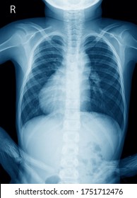
Chest X Ray 11 Year Old Stock Photo Edit Now 1751712476
X Ray Imaging For Covid 19 Patients
2
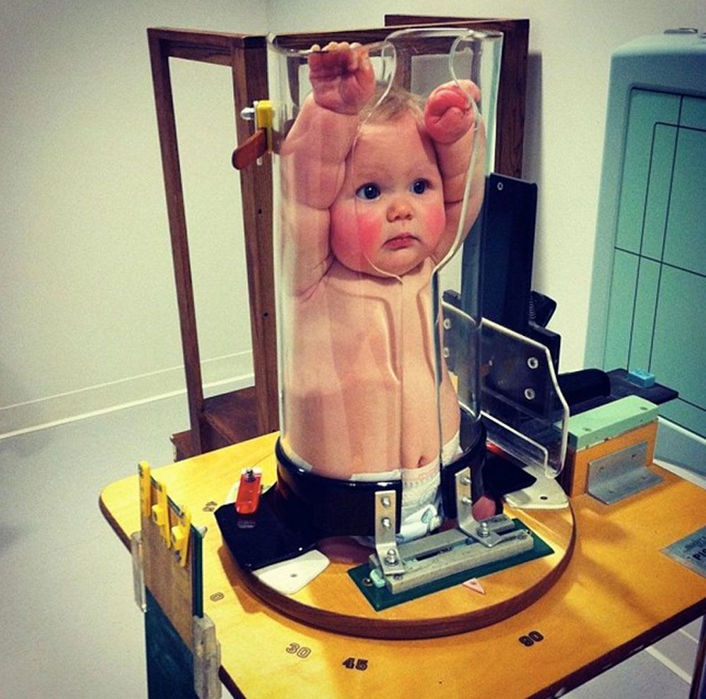
This Adorable Baby Is Squished Into A Tube For A Good Reason
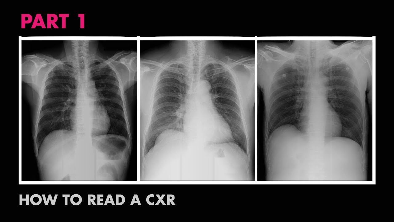
Assessment Of Cxr Positioning Views How To Read A Chest X Ray Part 4 Medzcool Youtube
2

Saber Sheath Trachea Radiology Case Radiopaedia Org Radiology Trachea Thoracic

Neonatal Radiography Part 1 Nomal Findings And The Basics Youtube

Pin By Eve Emmanuelle Roy Hebert On T I M Diagnostic Imaging Med Student Medical Field

4 Health Benefits And Risks Of X Rays No 1 Unique Dr Heben Human X Ray Walk In Clinic

Pediatric Chest Horizontal Beam Lateral View Radiology Reference Article Radiopaedia Org

Pediatric Chest Supine View Radiology Reference Article Radiopaedia Org